
5.6 Thyroid Eye Disease
Introductory Comments
Thyroid eye disease (TED) or thyroid orbitopathy is a common clinical examination case as patients are readily available, display numerous signs and may have potentially vision threatening complications. The thyroid eye examination is a tailored orbit examination. The orbit examination should be reviewed prior to reading this chapter.
Thyroid eye disease should be suspected in any patient who has lid retraction or proptosis, which can be observed on entering the room. Noting the presence of a thyroidectomy scar rapidly establishes the diagnosis. Once the diagnosis of thyroid eye disease is made, the purpose of the examination shifts to assessing for the activity and severity of the disease. Candidates should be familiar with clinical activity scoring and severity classifications.
Vision loss from thyroid eye disease may occur due to compressive optic neuropathy, exposure keratopathy, glaucoma, refractive errors, choroidal folds, and steroid induced cataract. Candidates will automatically fail the station if they do not assess for the presence of sight-threatening complications (compressive optic neuropathy, exposure keratopathy and glaucoma).
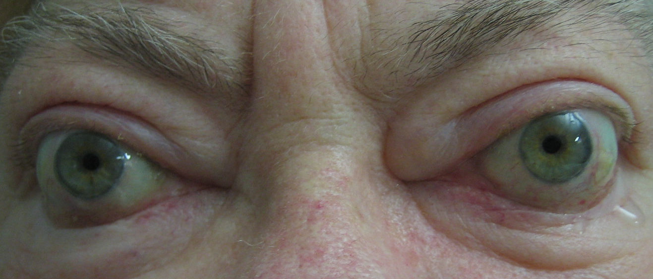
- Eyelid retraction
- Lid retraction should be suspected if the upper lid is level with or superior to the superior limbus. Look for superior scleral show and lateral flare. Patients may have a “thyroid stare” with infrequent blinking. Note that ptosis can occur in thyroid eye disease:
- Pseudo (eyelid retraction of the contralateral side)
- Real (aponeurotic, associated myasthenia gravis and CNIII palsy from crowding at the orbital apex)
- Lid retraction should be suspected if the upper lid is level with or superior to the superior limbus. Look for superior scleral show and lateral flare. Patients may have a “thyroid stare” with infrequent blinking. Note that ptosis can occur in thyroid eye disease:
- Soft tissue inflammation
- Erythema / oedema of the eyelids and conjunctiva (particularly look for injection over the insertion of rectus muscles and chemosis of the caruncle / plica)
- Proptosis
- TED is the most common cause of bilateral and unilateral axial proptosis
- Dystopia, strabismus
- Pupils (anisocoria)
- Systemic stigmata of thyroid disease
- Goitre / thyroidectomy scar
- Tremor / Clubbing (acropachy) / Onycholysis / Nicotine stains in smokers
- Tachycardia / Atrial Fibrillation
- Pretibial myxoedema
Hertel exophthalmometry should be undertaken to confirm the presence or absence of exophthalmos. If dystopia is noted estimate the horizontal and vertical displacement. Be careful when vertical strabismus is also present as this may simulate dystopia.
Previous lateral orbital decompression may be evident as a defect posterior to the lateral orbital rim. Assess for resistance to retropulsion.
- Pupils (RAPD)
- Red saturation
- Brightness saturation
- Visual fields to confrontation
- Colour vision testing
Absence of a RAPD and symmetry of perceived brightness / red saturation can indicate either normal optic nerve function or symmetrical bilateral optic neuropathy. If this is suspected, colour vision and visual fields should be formally checked.
Extraocular motility should be assessed. This can be limited by inflammation or fibrosis of extraocular muscles. The most common defects are hypotropia with limited elevation (due to inferior rectus restriction), followed by esotropia with limited abduction (due to medial rectus restriction). Limitations of depression (superior rectus restriction) and adduction (lateral rectus restriction) are less common. It is rare to have exotropia in thyroid eye disease - this suggests concomitant myasthenia gravis or complications from surgical decompression.
Lid lag
After testing extraocular motility with the usual “H” pattern, it is useful to check upgaze then downgaze. This is the time to observe for “lid lag” (when the upper lid lags behind the speed of the depression of the globe). Also observe whether the degree of lid retraction worsens on downgaze (retraction secondary to fibrotic levator palpebrae superioris) or conversely improves on downgaze (retraction secondary to a fibrotic inferior rectus and overaction of levator palpebrae superioris / superior rectus complex).
Myasthenia gravis is associated with Grave’s disease in approximately 10% of patients. See See Section 5.4 Ptosis.
The most common findings are:
- Superior limbic keratoconjunctivitis
- Look for superior conjunctival injection radiating back from the limbus and superior corneal PEEs. This is due to redundant conjunctiva sweeping onto the cornea with blinking
- Signs of corneal exposure (see above)
- Choroidal folds
- These are indicative of severe disease
- Optic nerve head
- Check for swelling (indicating acute compressive optic neuropathy), pallor (indicating chronic optic neuropathy) and glaucoma
Summary
After examining the patient and concluding the patient has TED, you must determine the:
- Severity (mild / moderate / severe / sight threatening)
- Clinical activity (active / inactive)
Severity should be assessed in all patients with thyroid eye disease. This determines the need for treatment. The European Group on Graves Orbitopathy (EUGOGO) have published a severity grading system [iv]:
- Mild (rarely require steroids)
- Dry eye is the most common symptom
- Moderate to severe
- Justifies immunosuppression (active) or surgery (inactive). 1 or more of the following:
- Diplopia in primary position
- Lid retraction ≥ 2mm
- Moderate or severe soft-tissue involvement
- Exophthalmos ≥ 3mm
- Diplopia in primary position
- Justifies immunosuppression (active) or surgery (inactive). 1 or more of the following:
- Severe sight-threatening thyroid eye disease (5%)
- Exposure keratopathy
- Optic nerve compression
- Exposure keratopathy
Bartalena L, Baldeschi L, Dickinson AJ, et al. Consensus statement of the European Group on Graves’ Orbitopathy (EUGOGO) on Management of Graves’ Orbitopathy. Thyroid 2008; 18(3):333-346.
Clinical activity should be assessed in all patients with thyroid eye disease. The natural history of thyroid eye disease usually follows “Rundle’s curve”, which is monophasic (becomes more active in the inflammatory stage, peaks at 6-24 months, then becomes less active in the fibrotic stage). Activity can be measured by several different methods – the candidate should be familiar with the more popular grading systems. Assessment of activity can guide method of treatment (a clinical activity score ≥ 4 / 10 indicates a greater likelihood of response to immunosuppression).
A. Clinical Activity Score (CAS, European) [v]
Mouritis MP, Prummel MF, Wiersinga WM and Koornneef L. Clinical activity score as a guide in the management of patients with Graves’ Ophthalmopathy. Clinical Endocrinology 1997; 47:9-14.
CAS ≥ 3 (first visit, or ≥ 4 for follow up visits) indicates active TED
Points 3 to 7 can be assessed by examination alone, the other points require history and prior information.
Pain
- Retrobulbar pain
- Pain on up / downgaze
Redness
- Eyelids
- Conjunctiva (Diffuse, > 1 quadrant)
Swelling
- Eyelids
- Conjunctiva (Chemosis)
- Caruncle
- ↑ Proptosis ≥ 2mm over 1 - 3 months
Dysfunction
- ↓ VA ≥ 1 line over 1 - 3 months
- ↓ Motility ≥ 5° over 1 - 3 months
On follow up visits the following can be assessed for 1 point each (with the total score then out of 10)
- Increase in measured proptosis > 2 mm over 1 - 3 months
- Decrease in eye movement limit of > 8 degrees over 1 - 3 months
- Decrease in visual acuity (2 Snellen chart lines) over 1 - 3 months
B. Visa Classification (Canadian) [vi] - Vision, Inflammation, Strabismus, Appearance
Dolman PJ and Rootman J. VISA Classification for Graves Orbitopathy. Ophthalmic Plastic and Reconstructive Surgery 2006; 22(5):319-324.
This classification system is more complex and grades thyroid disease according to these 4 factors. It is too complex and time-consuming for examinations and is more suited to long-term clinic management of thyroid eye disease patients
Occasionally candidates will be asked to discuss investigations. Most commonly they will be shown a copy of the patient’s CT orbits. Investigations that may be useful include:
Previous
5.5 Orbit
All rights reserved. No part of this publication which includes all images and diagrams may be reproduced, distributed, or transmitted in any form or by any means, including photocopying, recording, or other electronic or mechanical methods, without the prior written permission of the authors, except in the case of brief quotations embodied in critical reviews and certain other noncommercial uses permitted by copyright law.
Vitreoretinal Surgery Online
This open-source textbook provides step-by-step instructions for the full spectrum of vitreoretinal surgical procedures. An international collaboration from over 90 authors worldwide, this text is rich in high quality videos and illustrations.