
10 Equipment Stations
10.2 Keratometer
The keratometer measures anterior corneal curvature. This information is useful to:
- Study the pathological cornea (e.g. astigmatism, keratoconus)
- Fit contact lenses
- Calculate IOL power (K value in formulas calculating Intraocular Lens power)
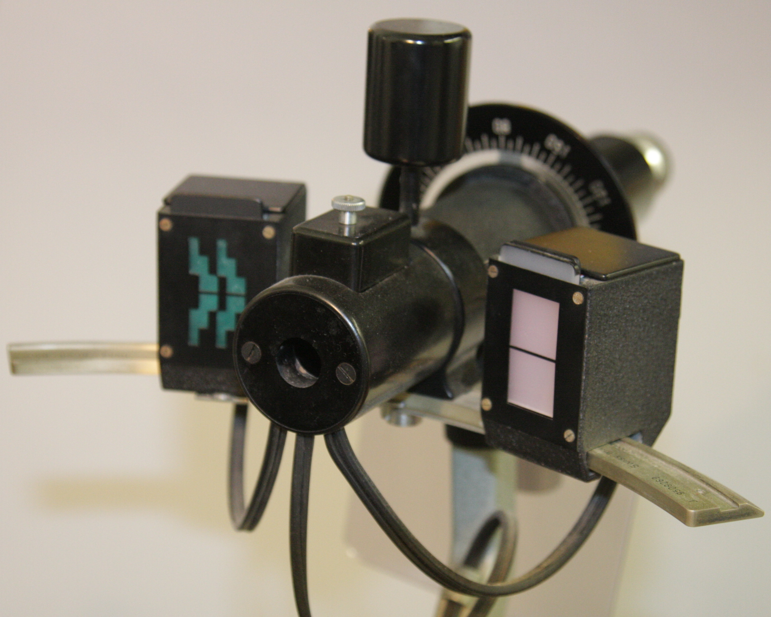
There are two main types: The von Helmholtz and Javal Shiotz. For both of them, the first steps are the same:
- Dim the room lights and turn the keratometer on
- Adjust your eyepieces. Turn the eyepiece towards the “plus” until the reticule is blurred. Turn it back the other way (towards the “minus”) until the reticule is just in focus
- Adjust the patient’s position (adjust the height of the chin rest and ensure that their forehead is against the band)
“Object” constant, “Image” adjusted
- Ask the patient to look straight into the keratometer and occlude the eye that is not being studied.
- Aim the keratometer until a reflection of the mires is seen on their cornea. Often finding this image is the most difficult part of the test. This can most easily be achieved by shining a pen torch in the keratometer and lining it up until a corneal reflection is seen (from the side, prior to looking in the keratometer). If the patient can’t tolerate the bright light, estimate the position with their eyelid closed
- Adjust the focus (forward / backward movement) until the 2 central circles completely overlap
- Adjust the axis of the keratometer (rotate it) until the “+”s and “-“s are aligned.
- Adjust the horizontal dial until the “+”s completely overlap. The reading on this dial is now the horizontal K reading (with the axis read off the keratometer)
- Adjust the vertical dial until the “-“s completely overlap. The reading on this dial is now the vertical K reading (with the axis read off the keratometer, normally 90° to the horizontal)
“Object” adjusted, “Image” constant
- Ask the patient to look towards the illuminated fixation point and occlude the eye that is not being studied
- Adjust the focus (forward / backward movement) with the joystick
- Adjust the axis of the keratometer (rotate it) until the central lines in the red and green mires align
- Adjust the dial until the red and green mires just touch. The reading on this dial is now the (horizontal) K reading (with the axis read off the keratometer)
- Turn the keratometer 90° and adjust the axis of the keratometer (rotate it) until the central lines in the red and green mires align. Since each “step” of the green “stepped” mire = 1 dioptre of corneal power, the amount of astigmatism is immediately obvious (if the mires just touch in one meridian and overlap by 2 steps in the meridian at 90° to this, there is 2 dioptres of corneal astigmatism). Again adjust the dial until the red and green mires just touch. The reading on this dial is now the (vertical) K reading (with the axis read off the keratometer, normally 90° to the previous)
-Keratometer.jpg)
-Keratometer-Mires.jpg)
Figure 10.2.2
von Helmholtz (Bausch & Lomb) Keratometer Mires
A: Adjust the focus until the 2 central circles completely overlap.
B: Adjust the axes until the “+”s and “-“s are aligned.
C: Adjust the horizontal dials until the “+”s completely overlap. The reading on this dial is now the horizontal K reading (with the axis read off the keratometer). Repeat in the vertical axis by aligning the “-“s.
Previous
10.1 Lensmeter
All rights reserved. No part of this publication which includes all images and diagrams may be reproduced, distributed, or transmitted in any form or by any means, including photocopying, recording, or other electronic or mechanical methods, without the prior written permission of the authors, except in the case of brief quotations embodied in critical reviews and certain other noncommercial uses permitted by copyright law.

Vitreoretinal Surgery Online
This open-source textbook provides step-by-step instructions for the full spectrum of vitreoretinal surgical procedures. An international collaboration from over 90 authors worldwide, this text is rich in high quality videos and illustrations.