
9 Investigations
9.1 Corneal Topography and Tomography
9.2 Confocal Microscopy
9.3 Optical Coherence Tomography - Macula
9.4 Optical Coherence Tomography Angiography (OCT-A)
9.5 Optical Coherence Tomography - Glaucoma
9.6 Optical Coherence Tomography – Anterior Segment
9.7 Fundus Autofluorescence Imaging
9.8 Fundus Angiography - Fluorescein
9.9 Fundus Angiography - Indocyanine Green
9.10 B-scan Ultrasonography & UBM
9.11 Electrophysiology
9.12 Automated Visual Fields
9.13 Neuroimaging
9.9 Fundus Angiography - Indocyanine Green
Indocyanine Green (ICG) Angiography, often used to complement Fundus Fluorescein Angiography (FFA), is a procedure used to acquire images of the choroidal vasculature. Indocyanine Green is a water-soluble dye which is almost entirely protein-bound (98%) which limits its diffusion out of the choroidal vasculature. It fluoresces in infrared light (790 – 805 nm) which has the ability to penetrate the retinal pigmented epithelium (RPE) and blood and allows for visualisation of the choroidal circulation. Following intravenous injection of ICG, the dye remains in the retinal and choroidal vessels and the fluorescence can be detected by fundus cameras or confocal scanning laser ophthalmoscopes. Images are captured at 1-minute intervals up until 5 minutes and then at 5 minute intervals up until 20 minutes.
Mild side effects of ICG include nausea, vomiting and pruritus. It should not be used in patients with an iodine allergy and in patents with liver disease or uraemia. ICG is classified as a Category C drug in pregnant women (have caused or may be suspected of causing, harmful effects on the human foetus or neonate without causing malformations - these effects may be reversible) and should be avoided.
Phases of ICG-Angiography:
i. Early Phase (First 1-minute Post Injection)
- Choroidal arteries shown
ii. Early Mid-phase (1 - 3 Minutes)
- Choroidal veins and retinal vessels seen clearly
iii. Late Mid-phase (3 - 15 Minutes)
- Structures fluorescing: Choroidal vessels fading but retinal vessels are still visible
iv. Late Phase (15 - 45 Minutes)
- Structures fluorescing: Gradual fading of diffuse hyperfluoresence
Normal Examples
.jpg)
.jpg)
- Polypoidal Choroidal Vasculopathy (PCV) – hyperfluorescent polyps
- Central Serous Chorioretinopathy (CSR)- mid-phase hyperfluorescence
- Choroidal inflammatory diseases such as birdshot choroidoretinopathy and APMPPE
- Choroidal Tumours such as choroidal haemangioma
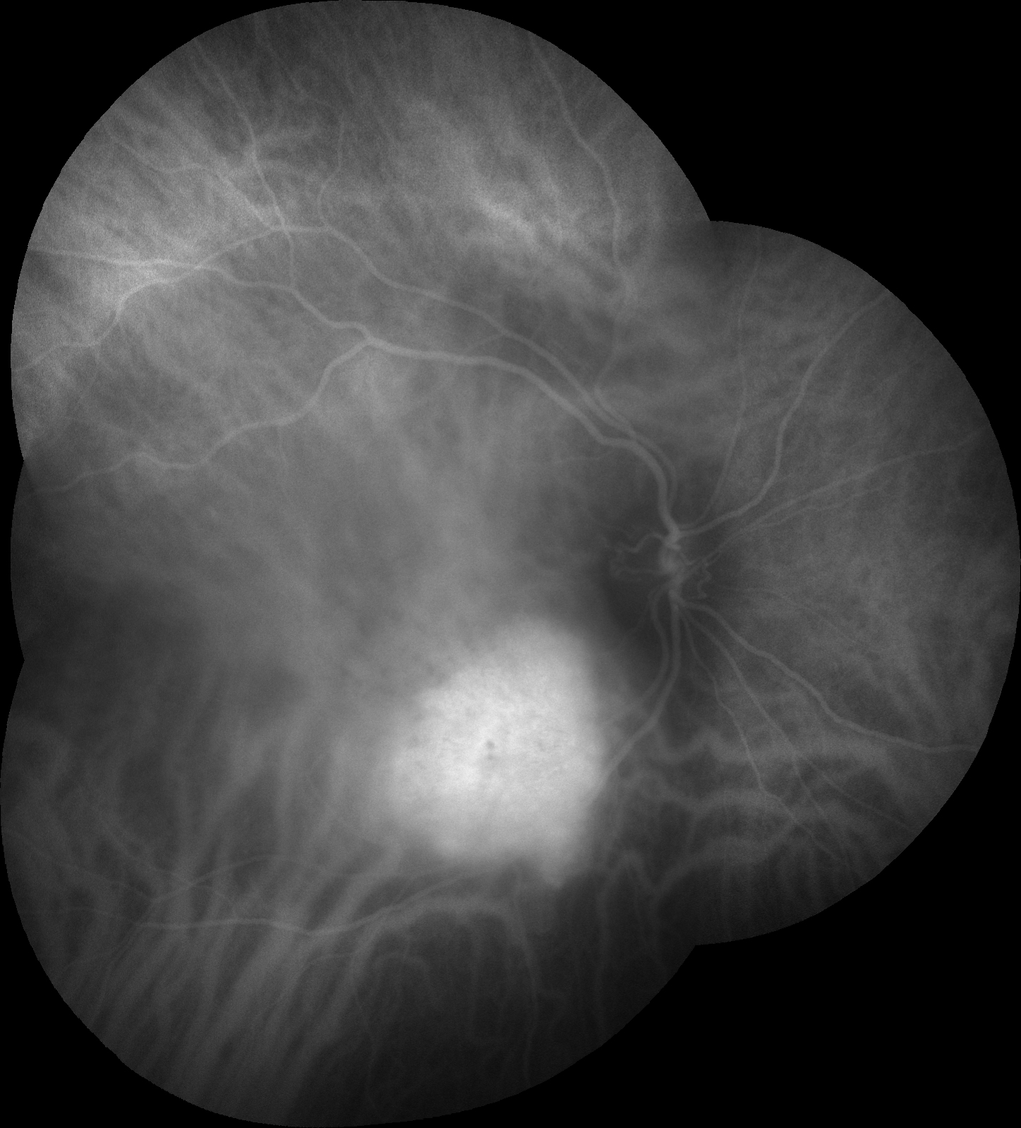
- Figure 9.9.3: Acute posterior multifocal placoid pigment epitheliopathy (APMPPE)
- Figure 9.9.4: Acute posterior multifocal placoid pigment epitheliopathy (APMPPE) (Late Phase)
- Figure 9.9.5: Choroidal Haemangioma
- Figure 9.9.6: Polypoidal Choroidal Vasculopathy (PCV)
- Figure 9.9.7: Ruptured Macroaneurysm
.jpg)
-(Late-Phase).jpg)
All rights reserved. No part of this publication which includes all images and diagrams may be reproduced, distributed, or transmitted in any form or by any means, including photocopying, recording, or other electronic or mechanical methods, without the prior written permission of the authors, except in the case of brief quotations embodied in critical reviews and certain other noncommercial uses permitted by copyright law.

Vitreoretinal Surgery Online
This open-source textbook provides step-by-step instructions for the full spectrum of vitreoretinal surgical procedures. An international collaboration from over 90 authors worldwide, this text is rich in high quality videos and illustrations.